JavaScript is disabled for your browser. Some features of this site may not work without it.
Supplementary information for the article: Virijević, K.; Živanović, M. N.; Nikolić, D.; Milivojević, N.; Pavić, J.; Morić, I.; Šenerović, L.; Dragačević, L.; Thurner, P. J.; Rufin, M.; Andriotis, O. G.; Ljujić, B.; Miletić Kovačević, M.; Papić, M.; Filipović, N. AI-Driven Optimization of PCL/PEG Electrospun Scaffolds for Enhanced In Vivo Wound Healing. ACS Appl. Mater. Interfaces 2024. https://doi.org/10.1021/acsami.4c03266.
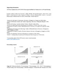
Figure S1. Publication Trends in “Electrospinning”, “Electrospinning + PCL + PEG”, “Electrospinning + Wound Healing” and “Electrospinning + Artificial Intelligence + Neural Network” Research (2001-2022) Table S1. Synonyms and Related Terms for Electrospinning in Research (2001-2022) Table S2. Input data structured for ANN – CSV data file Figure S2. Basic visualization of the dependence of output data (vertical axis) on individual input data (horizontal axis). Figure S3. Schematic representation of the neural network Figure S4. Graph of RMSE relation between training data set (blue line) and validation data set (orange line) depending on the number of neurons in the hidden layer Figure S5. Visual representation of ANN precision; the horizontal axis represents the percent of the real result, and the vertical axis represents the percent of ANN prediction; the training set (blue dots), prediction set (orange dots) Scheme S1. Electrospinning-Ready Polymer and Solvent Combinations. “Substanc...e” is PCL or PCL combined with PEG. The substance is dissolved in mass concentrations from 17 to 28% in CHCl3 or a combination of CHCl3 and DMF. Figure S6. Fiber Diameter Distribution Analysis of Electrospun-Derived Scaffold for Series 1: PCL in CHCl3. A) 17% B) 18% C) 19% D) 20% E) 21% F) 22% G) 23% H) 24% I) 25% J) 26% K) 27% Figure S7. Fiber Diameter Distribution Analysis of Electrospun-Derived Scaffold for Series 2: PCL in CHCl3:DMF=1:1. A) 17% B) 18% C) 19% D) 20% E) 21% F) 22% G) 23% H) 24% I) 25% J) 26% K) 27% Figure S8. Fiber Diameter Distribution Analysis of Electrospun-Derived Scaffold for Series 3: PCL in CHCl3:DMF=1:3. A) 17% B) 18% C) 19% Figure S9. Fiber Diameter Distribution Analysis of Electrospun-Derived Scaffold for Series 4: PCL in CHCl3:DMF=3:1. A) 17% B) 18% C) 19% D) 20% E) 21% Figure S10. Fiber Diameter Distribution Analysis of Electrospun-Derived Scaffold for Series 5: PCL:PEG=1:1 in CHCl3. A) 17% B) 18% C) 20% D) 21% E) 22% F) 24% G) 25% H) 26% I) 27% J) 28% Figure S11. Fiber Diameter Distribution Analysis of Electrospun-Derived Scaffold for Series 6: PCL:PEG=1:1 in CHCl3:DMF=1:1. A) 17% B) 18% C) 19% D) 20% E) 21% F) 22% G) 23% H) 24% I) 25% J) 26% K) 27% Figure S12. Fiber Diameter Distribution Analysis of Electrospun-Derived Scaffold for Series 7: PCL:PEG=1:1 in CHCl3:DMF=3:1. A) 17% B) 18% C) 19% D) 20% E) 21% F) 22% G) 23% H) 24% I) 25% Figure S13. Fiber Diameter Distribution Analysis of Electrospun-Derived Scaffold for Series 8: PCL:PEG=3:1 in CHCl3:DMF=1:1. A) 17% B) 19% C) 20% D) 21% E) 22% F) 24% G) 26% Figure S14. Fiber Diameter Distribution Analysis of Electrospun-Derived Scaffold for Series 9: PCL:PEG=3:1 in CHCl3:DMF=1:3. A) 17% B) 18% C) 19% D) 22% Figure S15. Fiber Diameter Distribution Analysis of Electrospun-Derived Scaffold for Series 10: PCL:PEG=3:1 in CHCl3:DMF=3:1. A) 17% B) 18% C) 19% D) 20% E) 21% F) 22% G) 23% H) 24% I) 25% J) 26% Figure S16. Fiber Diameter Distribution Analysis of Electrospun-Derived Scaffold for Series 15: PCL:PEG=1:3 in CHCl3:DMF=3:1. A) 17% B) 18% C) 19% D) 20% E) 21% F) 22% G) 23% H) 24% I) 25% Figure S17. Fiber Diameter Distribution Analysis of Electrospun-Derived Scaffold for Series 16: PCL:PEG=7:3 in CHCl3:DMF=7:3. A) 17% B) 18% C) 19% D) 20% E) 21% F) 22% G) 23% H) 24% I) 25% J) 26% K) 27% Figure S18. Fiber Diameter Distribution Analysis of Electrospun-Derived Scaffold for Series 17: PCL:PEG=3:1 in CHCl3. A) 17% B) 18% C) 19% D) 20% E) 21% F) 22% Figure S19. The action of scaffolds bearing antibiotics on selected strains of bacteria by disk diffusion method. Figure S20. Chick Embryo CAM Assay Procedure: A) Egg selection B) Egg disinfection with 10% of iodine solution C) Inoculation and preparation for scaffold insertion D) Egg’s incubation E) Daily monitoring of embryo development and possible contamination F) Sacrifice of treated embryos and fixation with 4% PFA G) CAM membrane preparation H) Image capture and evaluation of blood vessels
TY  - DATA
AU  - Virijević, Katarina
AU  - Živanović, Marko N.
AU  - Nikolić, Dalibor
AU  - Milivojević, Nevena
AU  - Pavić, Jelena
AU  - Morić, Ivana
AU  - Šenerović, Lidija
AU  - Dragačević, Luka
AU  - Thurner, Philipp J.
AU  - Rufin, Manuel
AU  - Andriotis, Orestis G.
AU  - Ljujić, Biljana
AU  - Miletić Kovačević, Marina
AU  - Papić, Miloš
AU  - Filipović, Nenad
PY  - 2024
UR  - http://intor.torlakinstitut.com/handle/123456789/873
AB  - Figure S1. Publication Trends in “Electrospinning”, “Electrospinning + PCL + PEG”, “Electrospinning + Wound Healing” and “Electrospinning + Artificial Intelligence + Neural Network” Research (2001-2022) Table S1. Synonyms and Related Terms for Electrospinning in Research (2001-2022) Table S2. Input data structured for ANN – CSV data file Figure S2. Basic visualization of the dependence of output data (vertical axis) on individual input data (horizontal axis). Figure S3. Schematic representation of the neural network Figure S4. Graph of RMSE relation between training data set (blue line) and validation data set (orange line) depending on the number of neurons in the hidden layer Figure S5. Visual representation of ANN precision; the horizontal axis represents the percent of the real result, and the vertical axis represents the percent of ANN prediction; the training set (blue dots), prediction set (orange dots) Scheme S1. Electrospinning-Ready Polymer and Solvent Combinations. “Substance” is PCL or PCL combined with PEG. The substance is dissolved in mass concentrations from 17 to 28% in CHCl3 or a combination of CHCl3 and DMF. Figure S6. Fiber Diameter Distribution Analysis of Electrospun-Derived Scaffold for Series 1: PCL in CHCl3. A) 17% B) 18% C) 19% D) 20% E) 21% F) 22% G) 23% H) 24% I) 25% J) 26% K) 27% Figure S7. Fiber Diameter Distribution Analysis of Electrospun-Derived Scaffold for Series 2: PCL in CHCl3:DMF=1:1. A) 17% B) 18% C) 19% D) 20% E) 21% F) 22% G) 23% H) 24% I) 25% J) 26% K) 27% Figure S8. Fiber Diameter Distribution Analysis of Electrospun-Derived Scaffold for Series 3: PCL in CHCl3:DMF=1:3. A) 17% B) 18% C) 19% Figure S9. Fiber Diameter Distribution Analysis of Electrospun-Derived Scaffold for Series 4: PCL in CHCl3:DMF=3:1. A) 17% B) 18% C) 19% D) 20% E) 21% Figure S10. Fiber Diameter Distribution Analysis of Electrospun-Derived Scaffold for Series 5: PCL:PEG=1:1 in CHCl3. A) 17% B) 18% C) 20% D) 21% E) 22% F) 24% G) 25% H) 26% I) 27% J) 28% Figure S11. Fiber Diameter Distribution Analysis of Electrospun-Derived Scaffold for Series 6: PCL:PEG=1:1 in CHCl3:DMF=1:1. A) 17% B) 18% C) 19% D) 20% E) 21% F) 22% G) 23% H) 24% I) 25% J) 26% K) 27% Figure S12. Fiber Diameter Distribution Analysis of Electrospun-Derived Scaffold for Series 7: PCL:PEG=1:1 in CHCl3:DMF=3:1. A) 17% B) 18% C) 19% D) 20% E) 21% F) 22% G) 23% H) 24% I) 25% Figure S13. Fiber Diameter Distribution Analysis of Electrospun-Derived Scaffold for Series 8: PCL:PEG=3:1 in CHCl3:DMF=1:1. A) 17% B) 19% C) 20% D) 21% E) 22% F) 24% G) 26% Figure S14. Fiber Diameter Distribution Analysis of Electrospun-Derived Scaffold for Series 9: PCL:PEG=3:1 in CHCl3:DMF=1:3. A) 17% B) 18% C) 19% D) 22% Figure S15. Fiber Diameter Distribution Analysis of Electrospun-Derived Scaffold for Series 10: PCL:PEG=3:1 in CHCl3:DMF=3:1. A) 17% B) 18% C) 19% D) 20% E) 21% F) 22% G) 23% H) 24% I) 25% J) 26% Figure S16. Fiber Diameter Distribution Analysis of Electrospun-Derived Scaffold for Series 15: PCL:PEG=1:3 in CHCl3:DMF=3:1. A) 17% B) 18% C) 19% D) 20% E) 21% F) 22% G) 23% H) 24% I) 25% Figure S17. Fiber Diameter Distribution Analysis of Electrospun-Derived Scaffold for Series 16: PCL:PEG=7:3 in CHCl3:DMF=7:3. A) 17% B) 18% C) 19% D) 20% E) 21% F) 22% G) 23% H) 24% I) 25% J) 26% K) 27% Figure S18. Fiber Diameter Distribution Analysis of Electrospun-Derived Scaffold for Series 17: PCL:PEG=3:1 in CHCl3. A) 17% B) 18% C) 19% D) 20% E) 21% F) 22% Figure S19. The action of scaffolds bearing antibiotics on selected strains of bacteria by disk diffusion method. Figure S20. Chick Embryo CAM Assay Procedure: A) Egg selection B) Egg disinfection with 10% of iodine solution C) Inoculation and preparation for scaffold insertion D) Egg’s incubation E) Daily monitoring of embryo development and possible contamination F) Sacrifice of treated embryos and fixation with 4% PFA G) CAM membrane preparation H) Image capture and evaluation of blood vessels
PB  - American Chemical Society
T2  - ACS Applied Materials & Interfaces
T1  - Supplementary information for the article: Virijević, K.; Živanović, M. N.; Nikolić, D.; Milivojević, N.; Pavić, J.; Morić, I.; Šenerović, L.; Dragačević, L.; Thurner, P. J.; Rufin, M.; Andriotis, O. G.; Ljujić, B.; Miletić Kovačević, M.; Papić, M.; Filipović, N. AI-Driven Optimization of PCL/PEG Electrospun Scaffolds for Enhanced In Vivo Wound Healing. ACS Appl. Mater. Interfaces 2024. https://doi.org/10.1021/acsami.4c03266.
DO  - doi.org/10.1021/acsami.4c03266
ER  -
@misc{
author = "Virijević, Katarina and Živanović, Marko N. and Nikolić, Dalibor and Milivojević, Nevena and Pavić, Jelena and Morić, Ivana and Šenerović, Lidija and Dragačević, Luka and Thurner, Philipp J. and Rufin, Manuel and Andriotis, Orestis G. and Ljujić, Biljana and Miletić Kovačević, Marina and Papić, Miloš and Filipović, Nenad",
year = "2024",
abstract = "Figure S1. Publication Trends in “Electrospinning”, “Electrospinning + PCL + PEG”, “Electrospinning + Wound Healing” and “Electrospinning + Artificial Intelligence + Neural Network” Research (2001-2022) Table S1. Synonyms and Related Terms for Electrospinning in Research (2001-2022) Table S2. Input data structured for ANN – CSV data file Figure S2. Basic visualization of the dependence of output data (vertical axis) on individual input data (horizontal axis). Figure S3. Schematic representation of the neural network Figure S4. Graph of RMSE relation between training data set (blue line) and validation data set (orange line) depending on the number of neurons in the hidden layer Figure S5. Visual representation of ANN precision; the horizontal axis represents the percent of the real result, and the vertical axis represents the percent of ANN prediction; the training set (blue dots), prediction set (orange dots) Scheme S1. Electrospinning-Ready Polymer and Solvent Combinations. “Substance” is PCL or PCL combined with PEG. The substance is dissolved in mass concentrations from 17 to 28% in CHCl3 or a combination of CHCl3 and DMF. Figure S6. Fiber Diameter Distribution Analysis of Electrospun-Derived Scaffold for Series 1: PCL in CHCl3. A) 17% B) 18% C) 19% D) 20% E) 21% F) 22% G) 23% H) 24% I) 25% J) 26% K) 27% Figure S7. Fiber Diameter Distribution Analysis of Electrospun-Derived Scaffold for Series 2: PCL in CHCl3:DMF=1:1. A) 17% B) 18% C) 19% D) 20% E) 21% F) 22% G) 23% H) 24% I) 25% J) 26% K) 27% Figure S8. Fiber Diameter Distribution Analysis of Electrospun-Derived Scaffold for Series 3: PCL in CHCl3:DMF=1:3. A) 17% B) 18% C) 19% Figure S9. Fiber Diameter Distribution Analysis of Electrospun-Derived Scaffold for Series 4: PCL in CHCl3:DMF=3:1. A) 17% B) 18% C) 19% D) 20% E) 21% Figure S10. Fiber Diameter Distribution Analysis of Electrospun-Derived Scaffold for Series 5: PCL:PEG=1:1 in CHCl3. A) 17% B) 18% C) 20% D) 21% E) 22% F) 24% G) 25% H) 26% I) 27% J) 28% Figure S11. Fiber Diameter Distribution Analysis of Electrospun-Derived Scaffold for Series 6: PCL:PEG=1:1 in CHCl3:DMF=1:1. A) 17% B) 18% C) 19% D) 20% E) 21% F) 22% G) 23% H) 24% I) 25% J) 26% K) 27% Figure S12. Fiber Diameter Distribution Analysis of Electrospun-Derived Scaffold for Series 7: PCL:PEG=1:1 in CHCl3:DMF=3:1. A) 17% B) 18% C) 19% D) 20% E) 21% F) 22% G) 23% H) 24% I) 25% Figure S13. Fiber Diameter Distribution Analysis of Electrospun-Derived Scaffold for Series 8: PCL:PEG=3:1 in CHCl3:DMF=1:1. A) 17% B) 19% C) 20% D) 21% E) 22% F) 24% G) 26% Figure S14. Fiber Diameter Distribution Analysis of Electrospun-Derived Scaffold for Series 9: PCL:PEG=3:1 in CHCl3:DMF=1:3. A) 17% B) 18% C) 19% D) 22% Figure S15. Fiber Diameter Distribution Analysis of Electrospun-Derived Scaffold for Series 10: PCL:PEG=3:1 in CHCl3:DMF=3:1. A) 17% B) 18% C) 19% D) 20% E) 21% F) 22% G) 23% H) 24% I) 25% J) 26% Figure S16. Fiber Diameter Distribution Analysis of Electrospun-Derived Scaffold for Series 15: PCL:PEG=1:3 in CHCl3:DMF=3:1. A) 17% B) 18% C) 19% D) 20% E) 21% F) 22% G) 23% H) 24% I) 25% Figure S17. Fiber Diameter Distribution Analysis of Electrospun-Derived Scaffold for Series 16: PCL:PEG=7:3 in CHCl3:DMF=7:3. A) 17% B) 18% C) 19% D) 20% E) 21% F) 22% G) 23% H) 24% I) 25% J) 26% K) 27% Figure S18. Fiber Diameter Distribution Analysis of Electrospun-Derived Scaffold for Series 17: PCL:PEG=3:1 in CHCl3. A) 17% B) 18% C) 19% D) 20% E) 21% F) 22% Figure S19. The action of scaffolds bearing antibiotics on selected strains of bacteria by disk diffusion method. Figure S20. Chick Embryo CAM Assay Procedure: A) Egg selection B) Egg disinfection with 10% of iodine solution C) Inoculation and preparation for scaffold insertion D) Egg’s incubation E) Daily monitoring of embryo development and possible contamination F) Sacrifice of treated embryos and fixation with 4% PFA G) CAM membrane preparation H) Image capture and evaluation of blood vessels",
publisher = "American Chemical Society",
journal = "ACS Applied Materials & Interfaces",
title = "Supplementary information for the article: Virijević, K.; Živanović, M. N.; Nikolić, D.; Milivojević, N.; Pavić, J.; Morić, I.; Šenerović, L.; Dragačević, L.; Thurner, P. J.; Rufin, M.; Andriotis, O. G.; Ljujić, B.; Miletić Kovačević, M.; Papić, M.; Filipović, N. AI-Driven Optimization of PCL/PEG Electrospun Scaffolds for Enhanced In Vivo Wound Healing. ACS Appl. Mater. Interfaces 2024. https://doi.org/10.1021/acsami.4c03266.",
doi = "doi.org/10.1021/acsami.4c03266"
}
Virijević, K., Živanović, M. N., Nikolić, D., Milivojević, N., Pavić, J., Morić, I., Šenerović, L., Dragačević, L., Thurner, P. J., Rufin, M., Andriotis, O. G., Ljujić, B., Miletić Kovačević, M., Papić, M.,& Filipović, N.. (2024). Supplementary information for the article: Virijević, K.; Živanović, M. N.; Nikolić, D.; Milivojević, N.; Pavić, J.; Morić, I.; Šenerović, L.; Dragačević, L.; Thurner, P. J.; Rufin, M.; Andriotis, O. G.; Ljujić, B.; Miletić Kovačević, M.; Papić, M.; Filipović, N. AI-Driven Optimization of PCL/PEG Electrospun Scaffolds for Enhanced In Vivo Wound Healing. ACS Appl. Mater. Interfaces 2024. https://doi.org/10.1021/acsami.4c03266.. in ACS Applied Materials & Interfaces
American Chemical Society..
https://doi.org/doi.org/10.1021/acsami.4c03266
Virijević K, Živanović MN, Nikolić D, Milivojević N, Pavić J, Morić I, Šenerović L, Dragačević L, Thurner PJ, Rufin M, Andriotis OG, Ljujić B, Miletić Kovačević M, Papić M, Filipović N. Supplementary information for the article: Virijević, K.; Živanović, M. N.; Nikolić, D.; Milivojević, N.; Pavić, J.; Morić, I.; Šenerović, L.; Dragačević, L.; Thurner, P. J.; Rufin, M.; Andriotis, O. G.; Ljujić, B.; Miletić Kovačević, M.; Papić, M.; Filipović, N. AI-Driven Optimization of PCL/PEG Electrospun Scaffolds for Enhanced In Vivo Wound Healing. ACS Appl. Mater. Interfaces 2024. https://doi.org/10.1021/acsami.4c03266.. in ACS Applied Materials & Interfaces. 2024;.
doi:doi.org/10.1021/acsami.4c03266 .
Virijević, Katarina, Živanović, Marko N., Nikolić, Dalibor, Milivojević, Nevena, Pavić, Jelena, Morić, Ivana, Šenerović, Lidija, Dragačević, Luka, Thurner, Philipp J., Rufin, Manuel, Andriotis, Orestis G., Ljujić, Biljana, Miletić Kovačević, Marina, Papić, Miloš, Filipović, Nenad, "Supplementary information for the article: Virijević, K.; Živanović, M. N.; Nikolić, D.; Milivojević, N.; Pavić, J.; Morić, I.; Šenerović, L.; Dragačević, L.; Thurner, P. J.; Rufin, M.; Andriotis, O. G.; Ljujić, B.; Miletić Kovačević, M.; Papić, M.; Filipović, N. AI-Driven Optimization of PCL/PEG Electrospun Scaffolds for Enhanced In Vivo Wound Healing. ACS Appl. Mater. Interfaces 2024. https://doi.org/10.1021/acsami.4c03266." in ACS Applied Materials & Interfaces (2024),
https://doi.org/doi.org/10.1021/acsami.4c03266 . .